
Figure 7. Incidence of SCaEs and corresponding DADs in current-clamped atrial myocytes from Ctl and cAF patients.
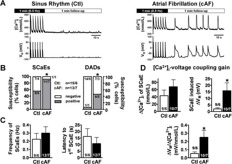
A, Representative recording of [Ca2+]i (Fluo-3) and corresponding membrane-voltage (Vm) oscillations (DADs/triggered APs) in a Ctl and a cAF-myocyte, respectively, following steady-state stimulation for 1-minute at 0.5 Hz. B, Enhanced susceptibility to spontaneous Ca2+-release events (SCaEs) and SCaE-induced DADs in cAF vs. Ctl. *P<0.05 vs. Ctl (Fisher’s exact test). C, Mean±SEM for frequency (left) and latency (right) of SCaEs. D, Mean±SEM amplitude of SCaEs (top left), magnitude of corresponding Vm-change (top right), and the calculated [Ca2+]i-membrane voltage coupling gain (bottom). *P<0.05 vs. corresponding means in Ctl. Numbers indicate myocytes/patients.