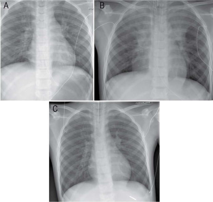
Figure 1A–C:
Portable post-intubation supine chest X-rays of a patient with pulmonary myiasis and eosinophilic pneumonia at (A) admission, (B) two days later and (C) four days after high-frequency oscillatory ventilation (HFOV). Note the worsening pneumonia with bilateral involvement and the improvement after HFOV.

