Figure 1.
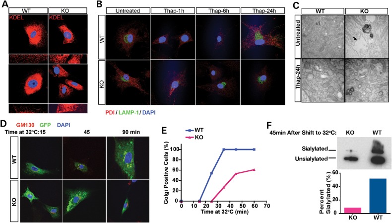
Plp2-deficient MEFs show dilated ER and defective ER-Golgi trafficking. (A) WT and KO MEFs were examined by confocal microscopy (400×) after staining with antibody specific for KDEL (red). Two representative fields of each genotype are shown with the peripheral ER of cells in each field shown at higher magnification (1000×) below. Nuclei were stained with DAPI (blue). (B) WT and KO MEFs were treated with Thap for the indicated times. ER and lysosome morphology were labeled with antibody specific for PDI (red) and LAMP-1 (green), respectively. Nuclei were stained with DAPI (blue). (C) Transmission electron microscopy of WT and KO MEFs with and without Thap treatment. Arrow points to the markedly dilated ER in the untreated KO MEFs. (D–F) Trafficking assay of WT and KO MEFs. (D) Immunofluorescence with antibody specific for the Golgi marker, GM130 (red). Nuclei were stained with DAPI (blue); ts-VSVG-GFP is shown in green. Note the co-localization of ts-VSVG-GFP with GM130 shown in yellow in the merged images. (E) Cells were counted under confocal microscopy. For each time point at least 100 cells were scored by an observer blinded to genotype, and the percentage of cells that show GFP positive in the Golgi is plotted on the y-axis. (F) Western blot. ts-VSVG was detected with antibody specific for GFP, and the unsialylated and sialylated forms reflect the ER form and Golgi-modified form of ts-VSVG-GFP, respectively.

