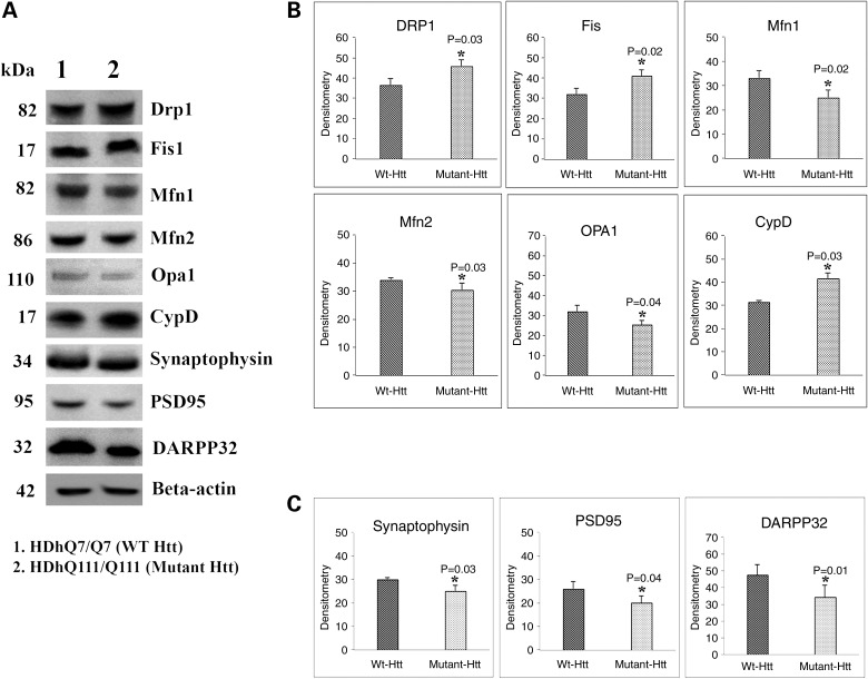
Figure 1.
Immunoblotting analysis of proteins in WT Htt (HDhQ7/Q7) and mutant Htt (HDhQ111/Q111) neurons. (A) Representative immunoblotting analysis of WT and mutant Htt neurons. (B) Quantitative densitometry analysis of mitochondrial dynamics and matrix proteins, Drp1, Fis1, Mfn1, Mfn2, Opa1 and CypD of WT Htt neurons relative to mutant Htt neurons. (C) Quantitative densitometry analysis of synaptic proteins synaptophysin, PSD95 and DARPP32 of mutant Htt neurons relative to WT Htt neurons. Significantly increased levels of fission proteins (Drp1 and Fis1) and significantly decreased levels of fusion and matrix proteins (Mfn1, Mfn2, Opa1 and CypD) in mutant Htt neurons relative to WT Htt neurons. Expression levels of synaptic protein levels synaptophysin and PSD95 and medium-spiny neuronal marker DARPP32 were significantly reduced in mutant Htt neurons.

