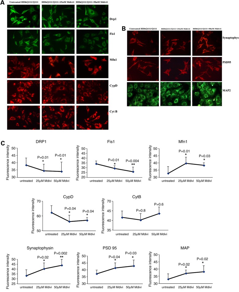
Figure 5.
Immunofluorescence analysis of proteins in Mdivil-treated and untreated mutant Htt neurons. (A) Representative images of Mdivi1-treated and untreated mutant Htt neurons from mitochondrial dynamics and matrix proteins. (B) Representative images of Mdivi1-treated and untreated mutant Htt neurons from synaptic proteins. (C) Quantitative immunofluorescence analysis of mitochondrial dynamics and matrix and synaptic proteins, Drp1, Fis1, Mfn1, Mfn2, Opa1, CypD and synaptophysin, PSD95 and MAP2 of mutant Htt (HDhQ111/Q111) neurons treated with Mdivi1 and untreated. The fission and matrix proteins were significantly decreased, and the fusion and ETC proteins were significantly increased upon treatment with Mdivil at 25 and 50 μm concentrations, indicating that Mdivi1 reduces mitochondrial fission activity and enhances fusion activity.

