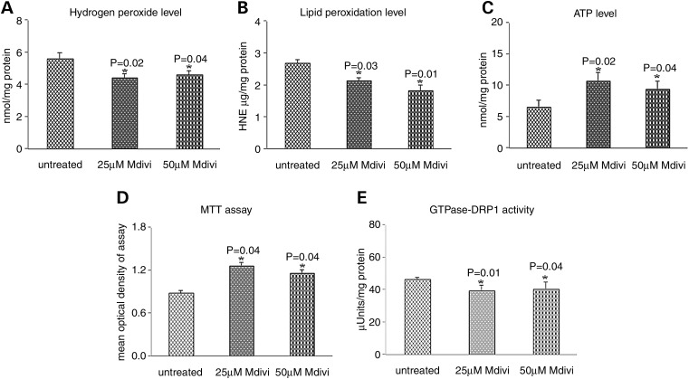
Figure 9.
Mitochondrial functional parameters in Mdivil-treated and untreated mutant Htt neurons. Mitochondrial function was assessed by measuring: (A) H2O2 production, (B) lipid peroxidation, (C) ATP levels, (D) cell viability and (E) GTPase Drp1 activity. Significantly decreased levels were found in the following parameters in mutant Htt neurons upon Mdivil treatment: H2O2 with Mdivil at 25 μm (P = 0.02) and 50 μm (P = 0.01) concentrations, and 4-hydroxy-2-nonenol with Mdivil at 25 μm (P = 0.03) and 50 μm (P = 0.01) concentrations. In contrast, significantly increased levels were found in the following parameters upon Mdivil treatment: ATP production at 25 μm (P = 0.02) and 50 μm (P = 0.04) concentrations, cell viability at 25 μm (P = 0.04) and 50 μm (P = 0.04) concentrations and GTPase Drp1 activity at 25 μm (P = 0.01) and 50 μm (P = 0.04) concentrations.

