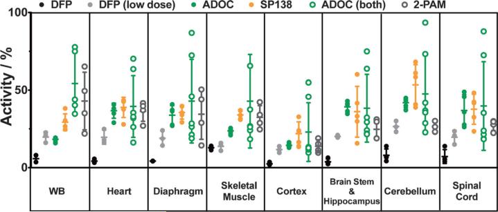
Figure 5.
Cholinesterase activity in mouse tissues after challenge with DFP (2). Animals were treated with 120 mgkg−1 total of ADOC (9) either 5 min after challenge (solid green circles) or 20 min before and 5 min (60 mgkg−1 per dose) after exposure (open green circles) to an otherwise lethal dose of 2 (3 mgkg−1). Another group of animals were fed 1 mg a day of SP138 (16, orange) for three days prior to exposure. All treated animals survived 24 h, after which tissues were collected and cholinesterase activity was measured. Activity from the treated animals was significantly higher than that from untreated control animals (i.e., 2 only), which expired within 90 min and are shown in black (two animals from each experiment). For comparison, three animals were treated with a lower dose of 2 (solid gray circles); they all survived for 24 h, albeit under visible distress, unlike the treated animals. Activity in tissues from across the BBB was also significantly higher than activity from a group of mice treated with pralidoxime (5) both 20 min prior to and 5 min post exposure (open gray circles). Activity is reported as the % activity in the samples relative to cumulative activity from tissue samples of control animals that were untreated and unchallenged. Activity measurements were repeated in triplicate and averaged; samples were normalized for protein content. Error bars show the standard deviation within each cohort of animals.

