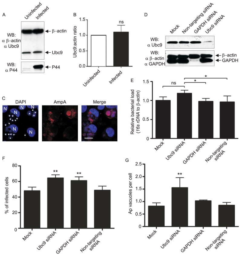
Fig. 8.
Ubc9 expression is not altered by A. phagocytophilum infection, but knockdown of Ubc9 results in a significantly increased bacterial load. A and B. A. phagocytophilum infection does not alter Ubc9 expression. A. Western-blotted whole cell lysates of uninfected and A. phagocytophilum infected cells were screened with β-actin and Ubc9 antibodies. B. Three pairs of infected and uninfected HL-60 lysates were examined in duplicate by Western blotting and the ratio of Ubc9 to β-actin analyzed by densitometry. “ns”, not significant. C. HEK-293 cells support A. phagocytophilum infection. HEK-293 cells that had been incubated with A. phagocytophilum were stained with DAPI (blue) to denote host cell nuclei (N) and bacteria (*). Cells were also stained with anti-AmpA (red) prior to visualization using LSCM. Scale bar, 10 μm. D–G. Effect of ubc9 knockdown on the A. phagocytophilum load in infected HEK-293 cells. HEK-293 cells were transfected with siRNA targeting ubc9 or gapdh, non-targeting siRNA, or transfection agent only (mock). At 72 h post transfection, the cells were boosted with a second siRNA transfection and infected with A. phagocytophilum. At 48 h post infection, the cells were resuspended and seeded on coverslips for immunofluorescent staining or collected for whole cell lysis and Western blotting analysis. D. Western blots demonstrating siRNA knockdown of Ubc9. Whole cell lysates from mock-, non-targeting siRNA-, gapdh siRNA-, and ubc9 siRNA-transfected cells were screened with antibodies targeting Ubc9 and GAPDH to confirm knockdown, and β-actin as a loading control. Blots are representative of more than four independent siRNA knockdown assays. E–G. The A. phagocytophilum load is modestly higher in host cells in which ubc9 has been knocked down. siRNA-treated and control host cells that had been infected with A. phagocytophilum were collected and processed for qPCR (E) or immunofluorescence microscopy (F and G). E. Relative loads of A. phagocytophilum 16S rRNA gene were normalized to human β-actin using the 2−ΔΔCT (Livak) method. Results shown are the means ± SD of triplicate samples and are representative of two independent experiments with similar results. Statistically significant (*P < 0.05) values relative to the ubc9 knockdown are indicated. F and G. Coverslips containing siRNA knockdown cells were processed for immunofluorescence and screened with anti-P44 to denote ApVs inside host cells. The mean ± standard deviations of percentages of infected HL-60 cells (F) and Ap vacuolar inclusions per HL-60 cell (G) were determined using immunofluorescence microscopy. Data presented are representative of two independent experiments. Statistically significant (*P < 0.05; **P < 0.005) values are indicated.

