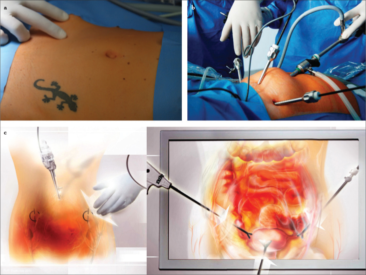
Figure 2.
a–c. Point of insertion from the outside (two thumbs medial of the anterior superior spine), at a 90° angle to the surface with penetration of all abdominal wall layers (a), trocar insertion site lateral to the plica umbilicalis lateralis (b), overview after insertion of the laparoscope and three ancillary trocars (c), graphical illustration of (a) and (b)

