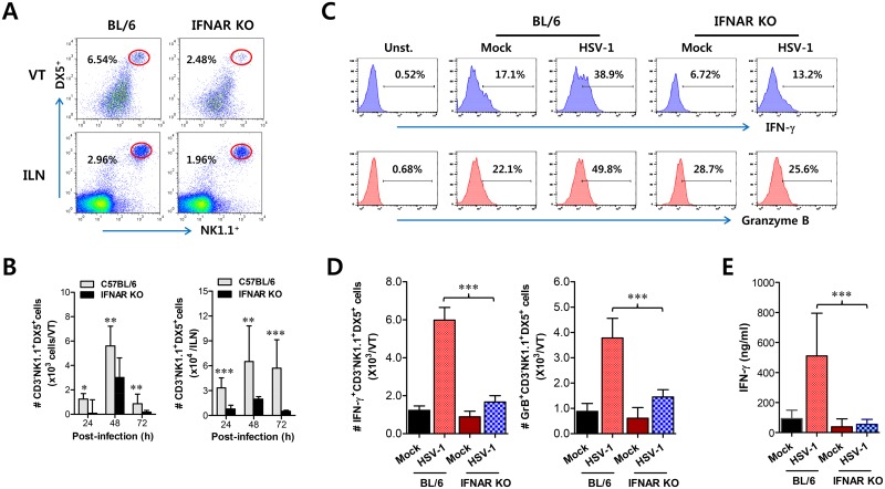
Fig 3. IFN-I signaling is involved in connected recruitment and activation of NK cells to CD11b+Ly-6Chi monocytes.
(A) NK cell infiltration of infected BL/6 or IFNAR KO mice. (B) Total accumulated NK cell number. Cells were prepared from vaginal tract (VT) and iliac LN (ILN) by collagenase digestion at 24, 48, and 72 h pi and used for analysis of NK cells. Values in dot-plots denote the average percentages of NK1.1+DX5+ NK cells derived from four independent samples after gating on CD3-negative cells at 48 h pi. (C) Frequency of IFN-γ or granzyme B-producing cells among NK cells. The production of IFN-γ and granzyme B by CD3−NK1.1+DX5+ NK cells was determined by intracellular staining after stimulation of vaginal NK cells with PMA plus ionomycin at 48 h pi. Values in histograms represent the average percentages of IFN-γ or granzyme B-producing cells among CD3−NK1.1+DX5+ NK cells. (D) Absolute number of IFN-γ or granzyme B-producing NK cells. Total number of IFN-γ or granzyme B-producing CD3−NK1.1+DX5+ NK cells in vaginal tract was enumerated by flow cytometric analysis using intracellular and surface staining at 48 h pi. (E) Secreted IFN-γ levels in vaginal tract. Secreted IFN-γ levels were determined by ELISA at 48 h pi using vaginal lavages. Data in bar chart represent the average ± SD of values derived from three individual experiments (n = 4–5). **, p<0.01; ***, p<0.001 compared with the levels of IFNAR KO mice.

