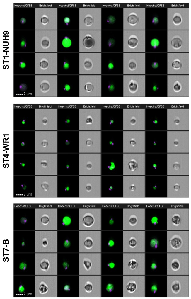
Fig 7. Image gallery of Blastocystis showing classical morphological forms at varying sizes in the viable populations.
Images were acquired using EDF setting of the imaging flow cytometer. Each cell is represented by two images: Hoechst-CFSE-staining composite and brightfield image. The nuclei were stained with Hoechst and the vacuole with CFSE. These round forms show a single nuclei located at the edge of the cell. These forms comprise more than half of the population in all the subtypes studied.

