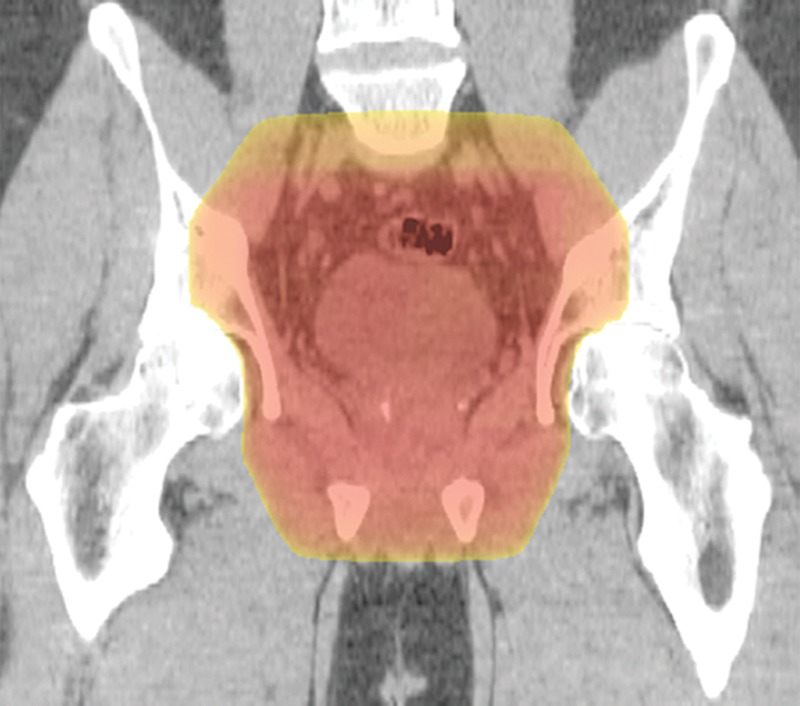
Figure 13a.

Example of a standard radiation treatment plan for rectal carcinoma. Coronal (a) and axial (b) CT images (obtained with prone patient position per radiation therapy standards) with superimposed radiation treatment plans show inclusion of the primary tumor bed and the presacral and internal iliac nodes. Higher-dose areas are more red; lower-dose areas are yellow. The external iliac nodes (not shown) were not included because the tumor did not invade anterior pelvic structures.
