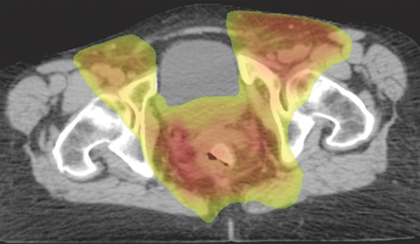
Figure 14b.

Representative example of a standard radiation treatment plan for anal carcinoma. Coronal (a) and axial (b) CT images show inclusion of the tumor, anus, perineum, and inguinal nodes. Higher-dose areas are more red, and lower-dose areas are more yellow-green. Note the attempt to spare radiation dose to the femoral heads.
