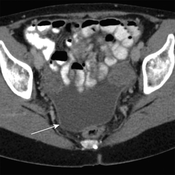
Figure 2b.

Key anatomic features of the rectum. (a, b) Axial computed tomographic (CT) images obtained at different levels in a patient with ascites show a peritoneal layer (arrow) covering the anterior and lateral upper rectum and a purely anterior peritoneal covering at the mid rectum. (c, d) In a different patient, axial T2-weighted magnetic resonance (MR) image (c) shows (from outside to inside) the mesorectal fascia (solid arrow), mesorectal fat, muscularis propria (arrowhead), submucosa, and mucosa (dashed arrow). Sagittal T2-weighted MR image (d) shows the relationship between the rectum and adjacent structures. The layers of the anus and rectum are also seen.
