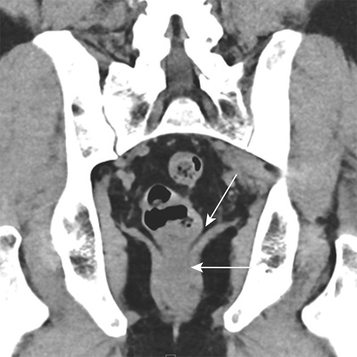
Figure 3a.

Key anatomic features of the anus. (a) Coronal CT image shows tapering of the mesorectal fascia around the anorectal junction at the pelvic floor. A thin fat plane is visible between the inner and outer sphincter muscles (arrows). (b) Axial T2-weighted MR image shows the internal sphincter (dashed arrow) and external sphincter (solid arrow). (c) Coronal T2-weighted MR image shows the confluence of the levator ani muscle (solid arrow) with the external anal sphincter (dashed arrow). The internal sphincter is contiguous with the rectal muscularis propria (arrowheads). (d) Sagittal CT image shows the relationship (anterior to posterior) of the bladder, seminal vesicles, rectum, and presacral fat. The anatomic rectosigmoid junction (bracket) is often defined at the sacral promontory or S3 to aid in treatment planning. The structure of the anal sphincter is poorly delineated on the CT image.
