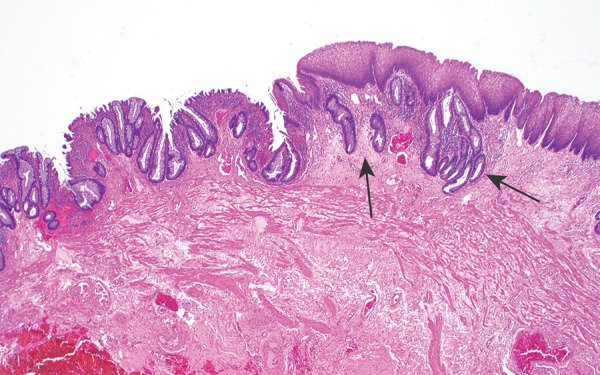
Figure 5.

Photomicrograph shows the anorectal junctional mucosa, with the transition from the columnar mucinous epithelium of the rectum (left) to the simple squamous epithelium of the anal canal (right); the transition is often somewhat irregular, and the rectal columnar epithelium can extend under the squamous mucosa for a short distance in this region (arrows). (Original magnification, ×40; hematoxylin-eosin [H-E] stain.)
