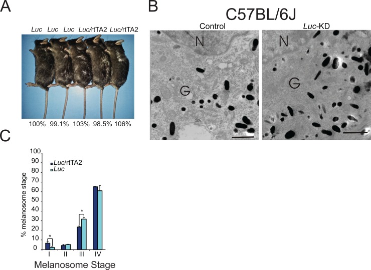
Fig 4. Expression of a control shRNA does not affect pigment accumulation or melanosome maturation.
(A) Coat color in Luc-knockdown mice and appropriate control mice was compared both visually (genotypes of each mouse are listed above each photo) and spectrophotometrically (the percentage value below each mouse corresponds to the absorbance at 492 nm for that particular mouse divided by the absorbance at 492 nm for its control littermate, which is set to 100%). (B) Fresh whole mouse skin was excised from Luc-knockdown mice and corresponding control mice using a four-mm round punch biopsy and fixed in Karnovsky’s fixative before electron microscopy analysis. Melanosomes within the Golgi area of the cell body were evaluated for maturation stage and ultra-structural morphology. Images are representative of 15 melanocytes from 2 mice per group. (C) The melanosomes were also quantified as described in Fig 3B. N = nucleus; G = Golgi area. Bar = 0.5 microns (Bar on inset = 1.5 microns). Graph: * = p ≤0.05. Bars = standard deviation (n = 2 mice per group, 15 melanocytes per mouse analyzed).

