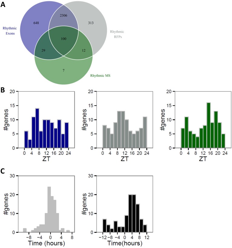
Fig. S3.
mRNA accumulation mainly drives protein rhythm. Proteins identified and quantified for at least eight time points in published MS data (14) were analyzed and compared with corresponding exonic and RFP reads obtained in our ribosome profiling data. Rhythmicity was assessed by performing a multiple linear regression for each relative time as described in ref. 14. Exonic, RFP, and MS signals were considered rhythmic for corrected P value (Benjamini–Hochberg method, ref. 82) below 0.25. (A) Venn diagram showing number of rhythmic proteins (green), mRNAs (blue), and rhythmic RFPs (gray). (B) Phase distributions of the 100 rhythmic proteins (green) encoded by rhythmic mRNA (blue) and RFP (gray). (C) Phase delays between mRNA and RFPs (Left) for group B show that phase of translation follows mRNA accumulation phase. Protein accumulation occurs mainly 4–6 h after the peak of RFP signal (Right).

