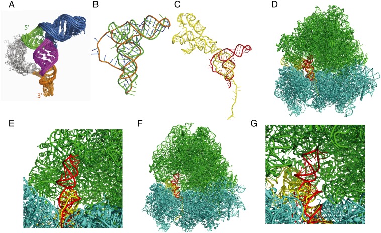
Fig. 7.
Structural model of the IAPV PKI domain. (A) Structural ensemble of the IAPV IRES, as determined by NMR/SAXS. The structural elements are colored as in Fig. 1. (B) Averaged structure of the IAPV PKI domain (orange) overlaid onto the structure of a Phe-tRNA (green). SLIII mimics the tRNA acceptor stem. (C) The IAPV PKI domain (red) superimposed onto the structure of the CrPV IGR IRES in the posttranslocated state (yellow) [Protein Data Bank (PDB) ID code 4D5Y)] (23). (D) The IAPV PKI domain (red) superimposed onto the structure of the CrPV IGR IRES bound in the A site of the yeast ribosome (PDB ID code 4V91) (20). The CrPV IRES (yellow), large ribosomal subunit RNA (green), and small ribosomal subunit RNA (cyan) are shown. (E) Zoom-in view of D, showing that SLIII (red) can be accommodated by occupying the space within the large ribosomal subunit normally occupied by the acceptor stem of a ribosomal A site tRNA (23). (F) The IAPV PKI domain (red) superimposed onto the structure of the CrPV IGR IRES bound in the P site of the O. cuniculus ribosome (PDB ID code 4V91) (20). (G) Zoom-in view of F.

