Fig. 1.
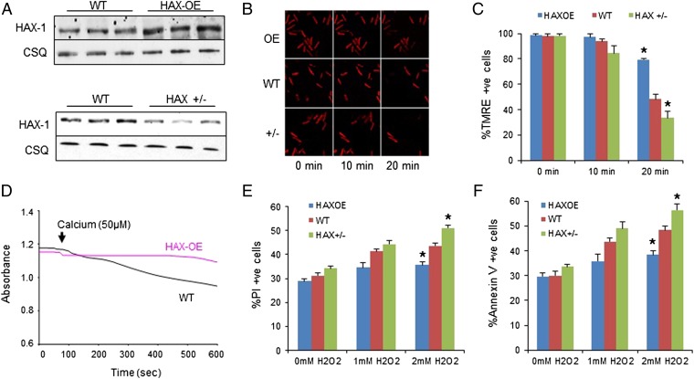
Overexpression of HAX-1 protects mitochondrial membrane integrity against oxidative stress and calcium overload, whereas heterozygous ablation of HAX-1 has opposite effects. (A) HAX-1 protein expression in HAX-OE and HAX+/− hearts. CSQ was used as loading control. (B and C) Isolated WT, HAX-OE, and HAX+/− cardiomyocytes were loaded with mitochondrial membrane potential fluorescent dye, TMRE. Upon 2 mM H2O2 administration for 20 min, membrane potential was diminished in all groups, but this effect was most pronounced in the heterozygous cells. HAX-OE exhibited more than 80% preserved TMRE signals. n = 7 hearts for HAX-OE, 16 hearts for WT, and 9 hearts for HAX+/− (>8 cells per heart); *P < 0.05 vs. WT at 20 min. (D) HAX-1 overexpressing mitochondria demonstrated resistance to swelling induced by 50 μM calcium. n = 3 hearts for each group. (E and F) HAX-1 overexpression in cardiomyocytes prevented cell death due to loss of plasma membrane integrity (E) and apoptosis (F) after 20 min of 2 mM H2O2 treatment, whereas HAX-1 heterozygous ablation increased cell death. n = 8 hearts for HAX-OE, 12 hearts for WT, and 13 hearts for HAX+/− (>10,000 cells per heart); *P < 0.05 vs. WT at 2 mM H2O2. Data are expressed as mean ± SEM.

