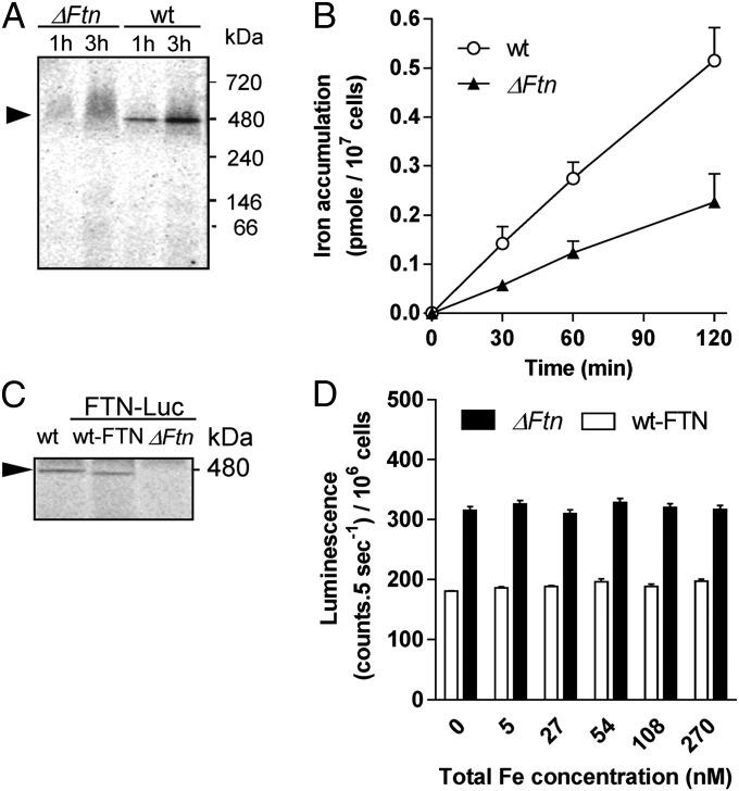
Fig. 2.
Functional analysis of ferritin in O. tauri. (A) Detection of ferritin on blue native PAGE. WT and ΔFtn cells were incubated in Mf medium with 1 µM 55Fe(III) citrate for 1 or 3 h and were washed twice with Mf medium. Protein extracts (25 µg per lane) were separated by blue native PAGE. Autoradiography of dried gel reveals one band at 480 kDa (black arrow) in WT but not ΔFtn protein extracts. (B) Iron uptake by WT and ΔFtn cells incubated in Mf medium with 1 µM 55Fe(III) EDTA (1:2). Values are means ± SD from three experiments. (C) Iron binding to ferritin in FTN-Luc (ΔFtn) knock-in and FTN-Luc (WT-FTN). 55Fe-labeled proteins (25 µg per lane) were analyzed in blue native PAGE as described in A. (D) Iron-dependent regulation of ferritin in FTN-Luc (ΔFtn) knock-in and FTN-Luc (WT-FTN). Cells acclimated for 7 d in 270nM Fe(III) EDTA were transferred to Aquil medium containing various amounts of total Fe. In vivo luminescence of reporter lines is plotted as a function of total Fe concentration. Data shown are means ± SD from five experiments (n = 5).

