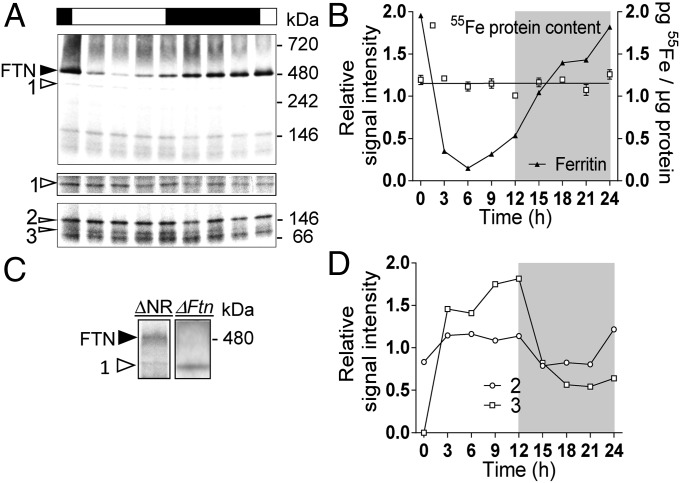
Fig. 4.
Detection of iron-binding proteins under a day/night cycle in stationary growth-phase cells of O. tauri. Proteins (25 µg per sample) from WT cells grown for 7 d with 1 µM 55Fe(III) citrate in Mf medium under a 12-h/12-h light/dark cycle were separated on blue native PAGE. (A) Autoradiography of radiolabeled proteins. Black and white boxes represent night and day, respectively; the two lower panels correspond to longer exposure time of the same gel. Arrowheads indicate the main iron-binding proteins, identified as ferritin (FTN) and putative NR (1), Fd-GOGAT (2), and NiR (3). See Table S1. (B) Quantitation of ferritin band intensity, normalized to the mean signal, reveals strong variation of radiolabeling over the day/night cycle. The 55Fe content of whole-protein extracts measured by liquid scintillation remained constant between the samples. (C) Autoradiography of Blue native PAGE (25 µg of protein in two different gels) of ΔFtn and ΔNR proteins from cells grown in Mf medium containing 1 µM 55Fe(III) citrate. Band 1 was absent from ΔNR cells but was present in ΔFtn cells which lack ferritin. (D) Quantitation of the main iron-binding proteins in A. The signal intensity is normalized to the mean signal.

