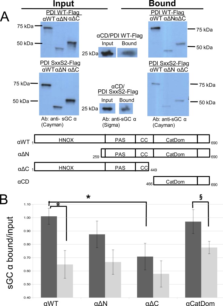
Fig 4. α sGC domains immunoprecipitation by Flag-tagged PDI-WT and Flag-PDI Ser-x-x-Ser2.
A. Representative Western blots of lysates (input) and immunoprecipitates (bound) using PDI-Flag and PDI-Flag mutant. COS-7 cells were transiently co-transfected with Flag-tagged PDI-WT or PDI Ser-x-x-Ser2 (PDI SxxS2) and sGC α domain constructs: αWT: full-length, αΔN: N-terminal truncation, αΔC: C-terminal truncation, α-CD: catalytic domain. Immunoprecipitation of lysates was performed using anti-Flag affinity resin as described in Material and Methods. Blots were probed with anti-sGC α Cayman and anti-sGC α Sigma. Bottom panel showing α constructs with domain boundaries labeled. B. Densitometry analysis of bound vs. input with ImageJ of three to five separate experiments. Black bars: PDI WT; gray bars: PDI SxxS2. Statistical analysis using Student's t-Test: α with PDI WT and α with PDI SxxS2, p = 0.01*; α with PDI WT and αΔC with PDI WT, p = 0.02*; and α-CatDom with PDI WT and α-CatDom with PDI SxxS2 = 0.06§.

