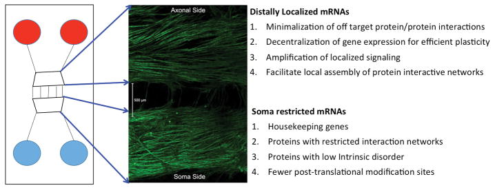
Figure 1. Use of Microfluidic Devices to elucidate properties of distally localized mRNAs.
The left panel shows a schematic of a microfluidic device while the middle panel shows an immunocytochemical image of DRG neurons in culture labeled with βIII-tubulin staining. DRG somas are found on the bottom side of the chamber and extend axons through the microfluidic barrier where they then elaborate extensive axonal arobrations on the axonal side. Properties of distally (e.g. axonal) localized mRNAs vs. those found restricted to cell bodies are listed on the right.

