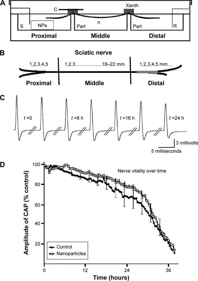
Figure 1.
Experimental setup and electrophysiology.
Notes: (A) Recording bath scheme. (B) Schematic representation of sciatic nerve’s proximal, middle, and distal regions. (C) Indicative evoked CAPs from whole nerve recordings. (D) Nerve vitality curve for CeO2.
Abbreviations: C, glass cover; CAP, compound action potential; n, perfusion chamber; NPs, nanoparticles; part, partition; R, recording chamber; S, stimulating chamber; Xanth, Xantopren; h, hours.

