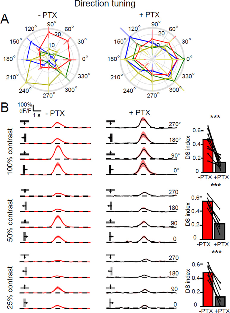
Figure 6. Directionally tuned responses require GABAergic signaling.
(A) Polar plots of calcium responses in T4 and T5 axon terminals to a moving dark bar, before and after application of PTX. Plots display the mean integrated responses to the bar moving in one of eight directions. Layers are color-coded: A = blue, B = green, C =red and D = yellow. Error bars are ±SEM. (B) Traces display mean responses. Shaded areas are ±SEM. The bar plots quantify the DS indices of these responses before and after PTX application to a moving dark bar at 100% contrast (upper plots). N = 10 flies; 50% contrast (middle plots), N = 6 flies; 25% contrast (lower plots), N = 6 flies. *** p<0.001, two tailed paired Student’s t-test. See also Figure S6.

