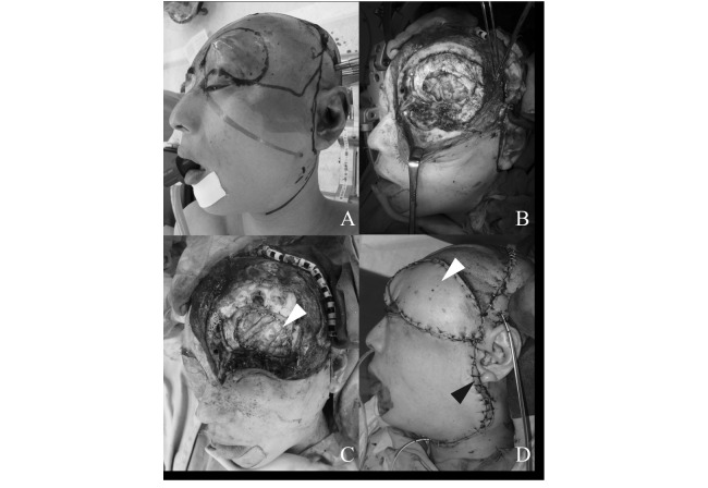
Fig. 3.
(A) Wide resection with a 2-cm margin from the edge of swelling was performed. (B) The left eye ball, dura mater, left frontal skull base, and the region of reddish tissue damaged by carbon ion radiotherapy were resected. (C) The dural defect was reconstructed using a free non-vascularized fascia lata graft (white arrow). (D) The soft tissue defect was reconstructed using a radial forearm flap (black arrow) and an anterolateral thigh flap (white arrow).

