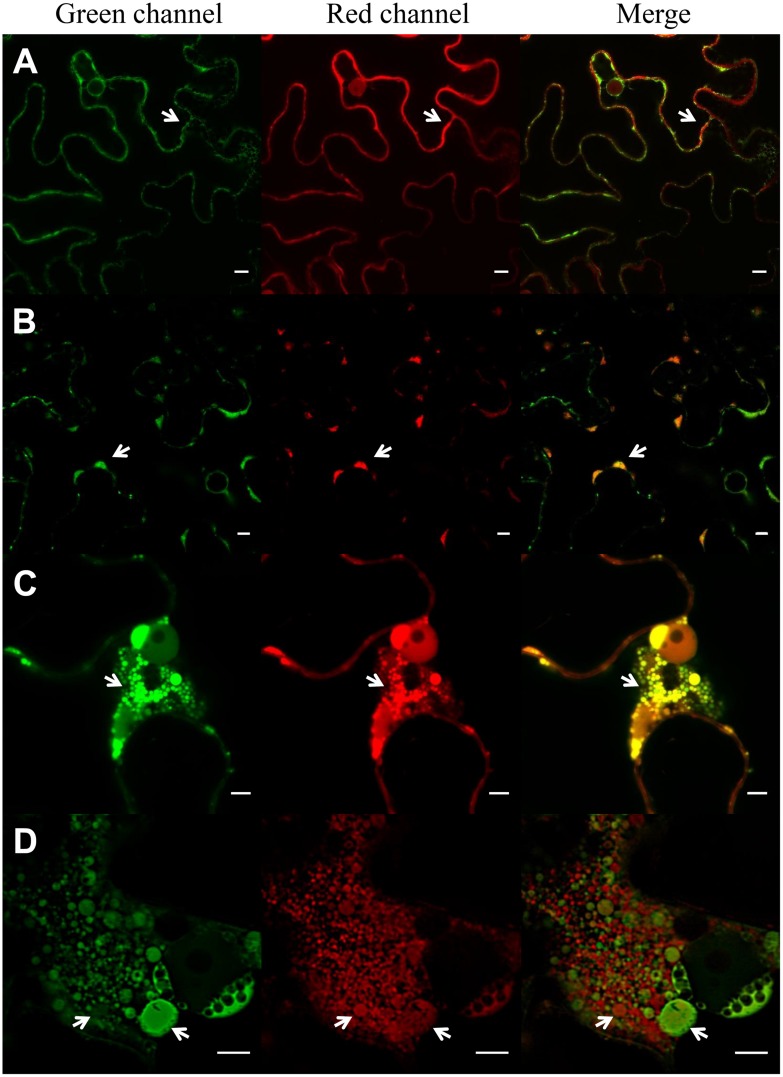
FIGURE 2.
Induction of protein bodies by expression of ELP. Expression of ER-GFP and sec-RFP (A), ER-RFP-TG2 and ER-GFP (B), ER-GFP, sec-RFP and ELP (C), ER-RFP-TG2, ER-GFP and ELP (D) in N. benthamiana leaf epidermal cells. ER-GFP shows the typical ER reticulated pattern (green channel, A), sec-RFP has an irregular pattern on the borders of the cell typical of apoplast (apo; red channel, A), no co-localization of ER-GFP and sec-RFP is observed in the merge panel. ER-RFP-TG2 (B) is mainly located in clusters (PB) in the borders of the cells (arrows), and the signal on the rest of the ER network is low. Co-localization of ER-RFP-TG2 and ER-GFP is observed mainly in these clusters (B, merge). ELP expression induces protein body formation (PB) in (C,D). Co-localization of ER-GFP and sec-RFP is observed in PB (C, merge) while RFP-TG2 did not entirely colocalize with ER-GFP (D, merge) in the nuclear region of the cell. Scale bars: 10 μm (A,B) and 5 μm (C,D).

