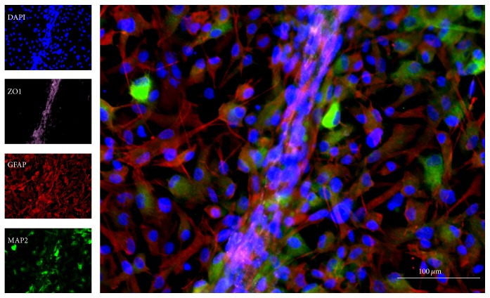
Figure 1.
In vitro modeling of the neurovascular environment using a novel method of coculturing human NSCs with human cerebral microvascular ECs, showing distinctive cytoarchitecture [11]. The composition of the transplant, including NSCs as well as other cellular and acellular compartments, can be investigated through paracrine and juxtacrine signaling within the neurovascular unit (NVU). Microtubule associate protein-2 (MAP2) is a neuronal marker; glial fibrillary acid protein (GFAP) is an astrocyte marker; zonula occludens 1 (ZO1) indicates the presence of tight junction protein; diamidino-2-phenylindole (DAPI) serves as a nuclear counterstain.

