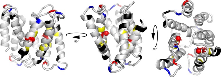
Figure 2.
Structure of homodimeric TPP+-bound EmrE with pH-sensitive residues highlighted. Basic residues are colored blue, and acidic residues are colored red. TPP+ is not shown. Note that Glu14, shown as sticks with the protonatable oxygen represented as a ball, is the only charged residue in the TM regions. Yellow residues (Ala10, Gly17, and Ser43) were used for the pKa fits and are located near the Glu14 active site. Black residues are more remote from the active site but also fit to the same two pKa values determined using the yellow residues close to Glu14 (Protein Data Bank accession no. 3B5D).

