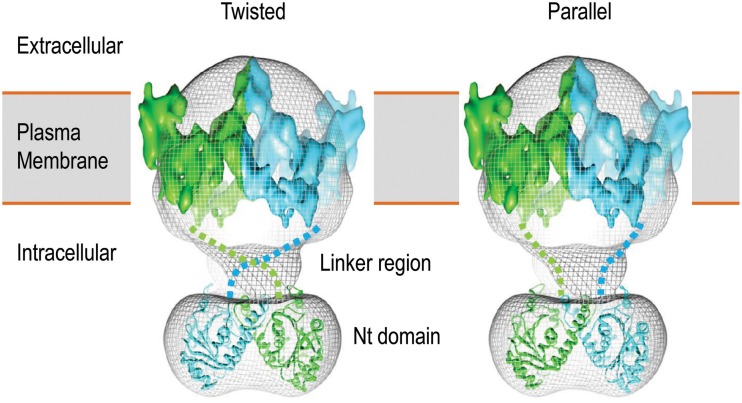
Figure 3.
3D structure model of human AE1 dimers obtained by cryo-EM. Shown here are two potential organizations of the AE1 dimers: twisted (left) and parallel (right). The two molecules of AE1 are shown in different color. The figures are modified from Jiang et al. (2013).

