
Figure 1:
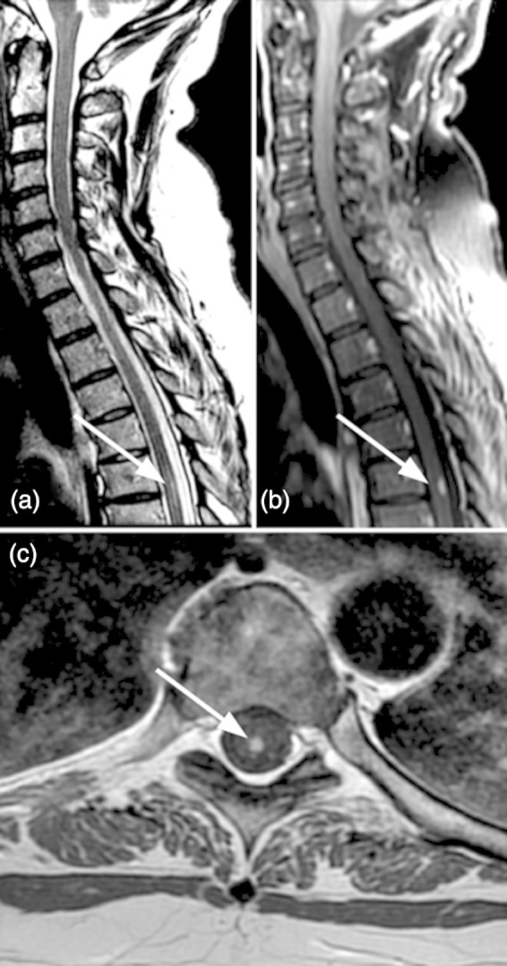
(a) Sagittal T2-weighted MRI shows a single posterior hyperintense demyelinating lesion of the cord at T5-6. (b) Sagittal CE T1 shows contrast enhancement of the lesion indicative of blood-spinal cord barrier disruption. (c): On axial CE T1 the lesion is located dorsomedially and limited to the posterior columns.