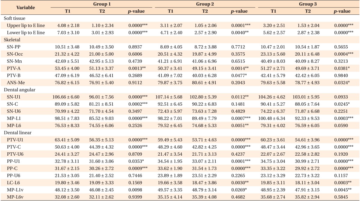
Table 3. Comparisons of the cephalometric measurements before treatment and after en masse retraction.
Values are presented as mean ± standard deviation.
Group 1, The C-lingual retractor group; group 2, the antero-posterior lingual retractor with parallel tube group; group 3, the antero-posterior lingual retractor with distally tipped tube group; T1, before treatment; T2, after en masse retraction.
The cephalometric landmarks for Tables 3 and 4 are the following: ANS-Me, lower anterior face height; LC-L6, mandibular lingual cortex to mandibular first molar centroid distance; MP-L1, mandibular plane to mandibular incisor angle; MP-L1v, mandibular plane to mandibular incisor tip distance; MP-L6, mandibular plane to mandibular first molar angle; MP-L6v, mandibular plane to mandibular first molar centroid distance; PP-C, palatal plane to maxillary canine tip distance; PP-U1, palatal plane to maxillary incisor tip distance; PP-U6, palatal plane to maxillary first molar centroid distance; PTV-A, pterygoid vertical plane to A point distance; PTV-B, pterygoid vertical plane to B point distance; PTV-C, pterygoid vertical plane to maxillary canine tip distance; PTV-U1, pterygoid vertical plane to maxillary incisor tip distance; PTV-U6, pterygoid vertical plane to maxillary first molar centroid distance; SN-PP, SN to palatal plane angle; SN-Mn, SN to mandibular plane angle; SN-Occ, SN-anatomic occlusal plane angle; SN-U1, SN to maxillary incisor angle; SN-U6, SN to maxillary first molar angle.
*p < 0.05, **p < 0.01, and ***p < 0.001, based on the paired t-test.
Refer Table 2 for definitions of the landmarks.

