. 2015 Nov 20;45(6):308–321. doi: 10.4041/kjod.2015.45.6.308

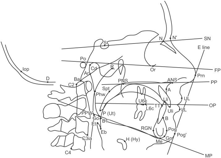
Figure 2. Diagrammatic representation of landmarks and reference lines. A, Subspinale; ANS, anterior nasal spine; Ar, articulare; B, supramentale; Ba, basion; BT', base of tongue; C2, tangent point on the dorsal surface of C2 vertebra to a line from C4; C3ia, C3 vertebra inferoanterior; C4, C4 vertebra inferoposterior; Cd, condylion; D, the deepest point in posterior cranial fossa; E line, Ricketts-E line; Eb, epiglottis base; Et, tip of epiglottis; FP, Frankfurt Plane; Gn, gnathion; Go, gonion; H (Hy), hyoidale; Iop: internal occipital protuberance; L1i, lower incisor tip; LL, lower lip; Me, menton; MP, mandibular plane; N, nasion; N', soft tissue nasion; OP, occlusal plane; Or, orbitale; P (Ut), soft-palate tip; Phw, posterior pharyngeal wall; PPW', posterior pharyngeal wall interseption; PNS, posterior nasal spine; Po, porion; Pog, pogonion; Pog', soft tissue pogonion; PP, palatal plane; Prn, nasal tip; R, roof of pharynx; RGN, retrognathion; S, sella; SN, S-N plane; Spt, tangent point on a line parallel to PNS-P on the dorsal surface of the soft palate at the maximum width; TT, tongue tip; U6c, maxillary first molar point; U1i, upper incisor tip; UL, upper lip.

Figure 2