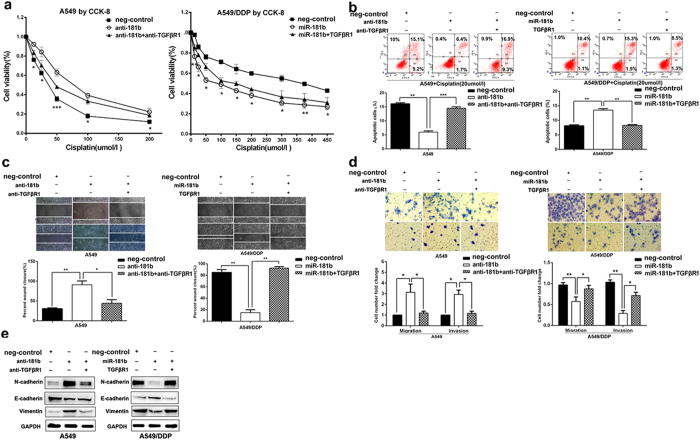
Figure 5. TGFβR1 has a critical role in miR-181b-mediated cell growth, chemosensitivity to DDP and metastasis of NSCLC cells.
(a) CCK analysis of the IC50 values of DDP in A549/miR-181b inhibitors, A549/miR-181b inhibitors/si-TGFβR1, A549/DDP/miR-181b or A549/DDP/miR-181b/TGFβR1. (b) Flow cytometric analysis of apoptosis in A549/miR-181b inhibitors, A549/miR-181b inhibitors/si-TGFβR1, A549/DDP/miR-181b or A549/DDP/miR-181b/TGFβR1 with DDP treatment. (c,d) Scratch, migration, invasion assay in A549/miR-181b inhibitors, A549/miR-181b inhibitors/si-TGFβR1, A549/DDP/miR-181b or A549/DDP/miR-181b/TGFβR1. (e) Western blot detection of E-cadherin, Vimentin, N-cadherin protein expression in A549/miR-181b inhibitors, A549/miR-181b inhibitors/si-TGFβR1, A549/DDP/miR-181b or A549/DDP/miR-181b/ TGFβR1. GAPDH was used as an internal control. *P < 0.05; **P < 0.01; ***P < 0.001.

