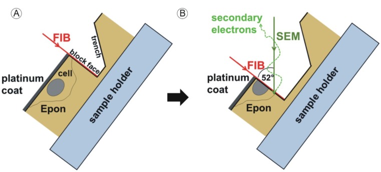
Figure 1.
Sample manipulation by the focused ion beam (FIB) and image acquisition with the scanning electron microscope (SEM) beam. Sample preparation is described in [39]. Embedded HCMV infected fibroblasts are located directly under the surface of the approximately 1 mm high Epon block. The sample is coated with a thin platinum layer prior to mounting into the dual beam microscope. The area of interest is chosen at 10 kV acceleration voltage. (A) The sample is then tilted to 52° and a trench is FIB-milled into the Epon block, generating a new surface (block face); (B) Removal of the next thin layer (“slice”) of the sample generates a new block face. After every “slice” step the generated block face is imaged by the scanning electron beam (“view”). The “slice and view” cycle is repeated to gain a three-dimensional dataset of the volume of interest. Reprinted from [38] Figure 1 with kind permission from Springer Science+Business Media.

