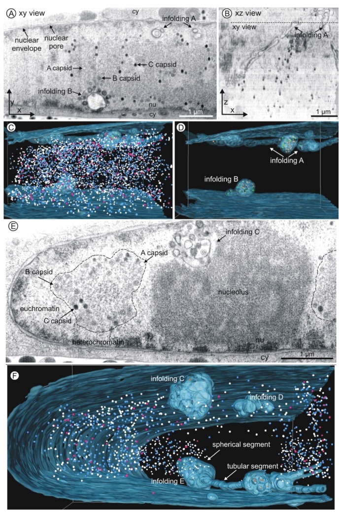
Figure 3.
FIB/SEM tomography of an HCMV infected nucleus five days post infection. (A–D) Volume 1; (E–F) Volume 2; (A,E) Single FIB/SEM micrographs. High contrast allows identification of membranes and nuclear capsids. Nuclear membrane structures (infoldings) are visible close to the nuclear envelope (see also Movies S1 and S2). nu nucleoplasm, cy cytoplasm. Dimensions of each volume: 6.4 µm × 5.5 µm × 5 µm; (B) Xz view of the tomogram of volume 1. The dashed line marks the position of the FIB/SEM image shown in (A). (C,D,F) Three-dimensional reconstructions of the inner nuclear membrane with infoldings (blue, semi-transparent) and capsids (nucleoplasm: A capsids pink; B capsids blue; C capsids white; infoldings: A capsids purple; B capsids orange; C capsids green). See also Movies S3 and S4. (C and D) Volume 1 contains 4160 capsids. (E) The dashed line marks a replication center. (F) Infolding D has no connection to the inner nuclear membrane. Infolding E consists of tubular and spherical segments. Volume 2 contains 1338 capsids.

