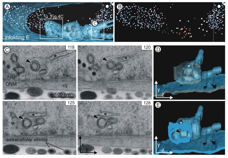
Figure 5.
Infolding E. (A) Two large spherical segments (diameters 1 µm) are connected by a tubular segment (diameter ~180 nm). A smaller spherical segment at the front branches out into two tubular segments. The boxes mark the position of the details depicted in Figure 4C and Figure 5C–E; (B) Presentation of the capsids alone shows the cloud-like structure of the nucleoplasmic capsids; (C) A 3rd order infolding (arrowhead) is visible in a sequence of more than ten images (see Movie S4). The images also show an invagination within the tubular segment (asterisk). Its connection with the nucleoplasm is evident in image 128. INM inner nuclear membrane, ONM outer nuclear membrane, nu nucleoplasm, cy cytoplasm; (D,E) The yz view of the three-dimensional model shows the opening of the invagination (asterisk). The model is depicted without capsids.

