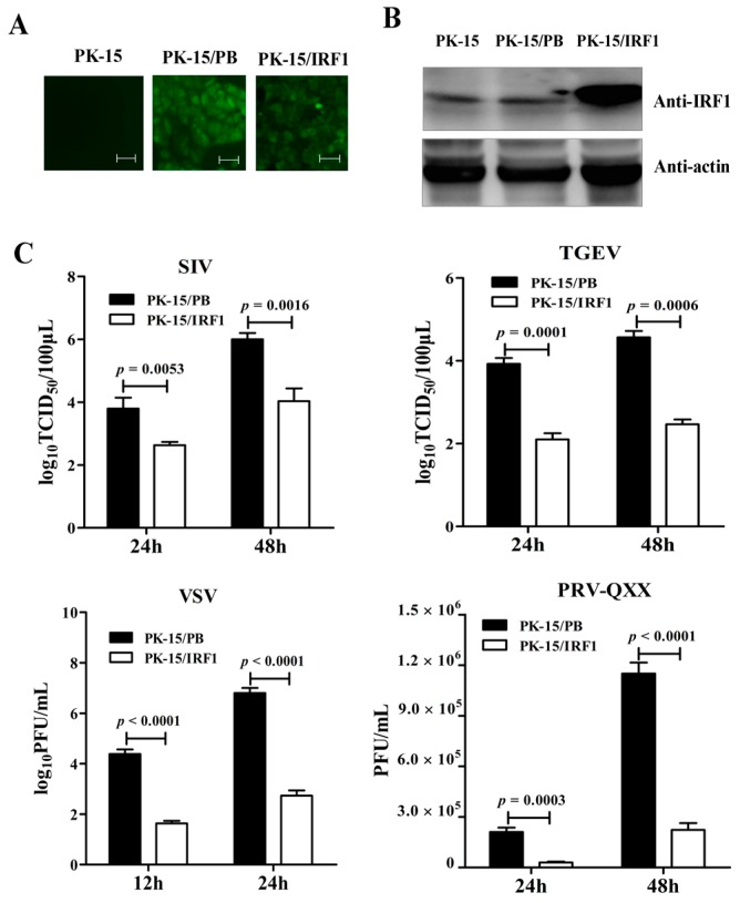
Figure 2.
Overexpression of poIRF1 potently inhibits virus replication. (A) PK-15 cells stably expressing poIRF1 were established by PB transposon systems. After puromycin treatment, the single puromycin-resistant and GFP-positive cell clones, designated as PK-15/IRF1 and PK-15/PB, were captured on a fluorescence microscope; scale bar, 100 µm; (B) Detection of poIRF1 protein by Western blot. Protein lysates from PK-15, PK-15/PB and PK-15/IRF1 were denatured in SDS sample buffer. Denatured lysates were analyzed by SDS-PAGE followed by Western blot using antibodies detecting poIRF1, as indicated. β-actin was used as a loading control; (C) The impact of poIRF1 overexpression on viral replication. PK-15 cells stably expressing poIRF1 or with vector alone were incubated with indicated viruses for 24 h and 48 h before supernatants were harvested. The viral titers in supernatants were measured with TCID50 assay or plaque assay.

