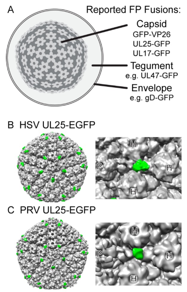
Figure 2.
Structure of the Alpha Herpesvirus particle. (A) Virions are composed of an icosahedral capsid containing the viral genome, a complex and pleomorphic tegument, and pleomorphic envelope. See Table 1 for a complete list and references of viral structural proteins reported to tolerate fluorescent protein fusion. (B,C) Enhanced green fluorescent protein (EGFP) fusions to viral capsid protein UL25 can be detected by cryo electron microscopy in herpes simplex virus 1 (HSV-1) and pseudorabies virus (PRV). Five copies of UL25 are bound to each penton. Due to the elongated conformation of UL25 [98], the EGFP moiety (green) appears distal to the penton (P), between hexons (H). The EGFP density thus serves as a convenient mass tag to aid in determining the location of particular viral proteins on the capsid. (B) A 13.7 Å icosahedral reconstruction of an HSV-1 nuclear C-capsid, rendered from EMDB-1904, originally published by Conway et al. [99]. (C) A 23.2 Å icosahedral reconstruction of a PRV nuclear C-capsid, rendered from EMDB-5657, originally published by Homa et al. [100].

