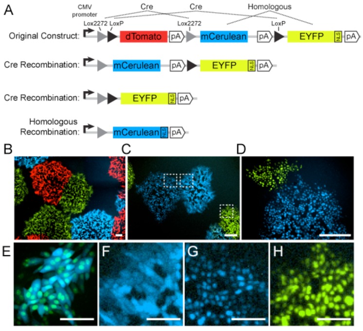
Figure 3.
Recombination between homologous fluorescent proteins. (A) Herpes simplex virus 1 (HSV-1) 17syn+ strain containing the Brainbow 1.0L cassette inserted into the UL37/UL38 intergenic region. The original construct expresses dTomato (red). Upon co-expression with Cre recombinase, site-specific recombination at Lox sites produces progeny virus genomes that express either mCerulean (cyan) or enhanced yellow fluorescent protein (EYFP; depicted in green) fused to a nuclear localization signal (NLS). Due to the high sequence similarity between mCerulean and EYFP, non-Cre homologous recombination can transfer the NLS to mCerulean, producing an unexpected phenotype. (B) HSV-1 plaques expressing dTomato, mCerulean, or EYFP-NLS. (C) HSV-1 plaques expressing mCerulean, EYFP-NLS, or unexpectedly, mCerulean-NLS. Boxed regions are magnified in panels F-H. (D) HSV-1 plaques expressing EYFP-NLS, or unexpectedly, mCerulean-NLS. Scale bars in panels B-D are 250 µm. (E) Cells co-infected with HSV-1 expressing mCerulean and EYFP-NLS, illustrating the distinction between cytosolic and nuclear localization. (F) Cells infected with HSV-1 expressing cytosolic mCerulean, as expected. (G) Cells infected with HSV-1 unexpectedly expressing mCerulean-NLS. (H) Cells infected with HSV-1 expressing EYFP-NLS, as expected. Scale bars in panels E–H are 100 µm.

