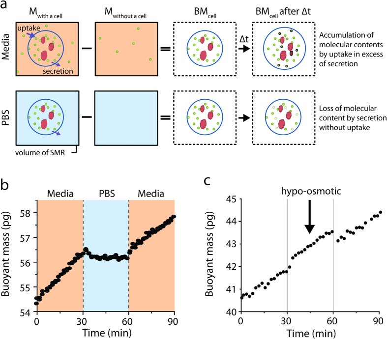
Figure 1. The SMR measures instantaneous accumulation of molecular contents in a single cell.
(a) The difference in SMR mass (black box) with or without a cell is the buoyant mass of a cell. Since ion concentration inside and outside the cell is approximately the same, the buoyant mass of a cell is determined by molecular contents such as small molecules (green circle–metabolites or amino-acids) or macromolecules (red globule–proteins, lipids, or nucleic-acids). In media, cellular uptake exceeds secretion thus molecular contents accumulate over time (dark green and red indicate newly acquired molecules). In PBS when all environmental nutrients are absent, cells only secrete molecules and molecular contents decrease (dim green indicates the lost molecules). (b) A cell’s buoyant mass is measured while its surrounding is rapidly switched from media to PBS (t = 30 minutes) and back to media (t = 60 minutes). The orange and blue background mark culture media and PBS, respectively, as in (a). The transient buoyant mass change at the transition results from the density and osmolarity mismatch between the media and PBS. The responses from additional cells are shown in Supplementary Fig. 3. (c) Changing the density and osmolarity can offset the cell’s buoyant mass but the mass accumulation rate remains unchanged. Hypo-osmotic condition was achieved by diluting standard RPMI media to 20 mM lower osmolarity with deionized water.

