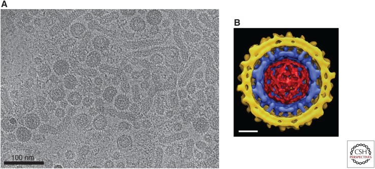
Figure 4.
The hepatitis B virus (HBV) virion. (A) Cryoelectron micrograph (cryo-EM) of virions isolated from chronically infected patients showing virions (Dane particles) containing spherical envelopes and virions with elongated appendages. Also shown are spherical and filamentous subviral particles (provided by Dr. Stefan Seitz). (B) Composite cryoelectron microscopy model of an HBV virion comprising the viral lipid envelope with hepatitis B surface antigen (HBsAg) protrusions (yellow), the icosahedral capsid (blue), and the enclosed double-stranded DNA (dsDNA) (red) (Dryden et al. 2006). The capsid displays icosahedral symmetry with protrusions representing the 4-helix bundles of the hepatitis B core antigen (HBcAg) dimers. Protrusions from the envelope are dimers of predominantly the small (S) form of the envelope protein, S-HBsAg. The viral envelope interacts with the spike tips of the capsid but is not arranged with icosahedral symmetry. Scale bar, 10 nm

