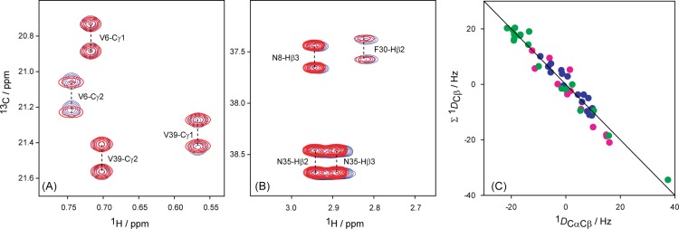
Figure 2.
Examples of spectral quality used for deriving 1DCβCγ couplings. (A) Small section of the methyl region of the 1H–13C HSQC spectrum of wild-type GB3 in Pf1 medium, recorded with regular (non-CT) 13C evolution (red), superimposed on the corresponding spectrum recorded under isotropic conditions (blue). (B) Small region of the 1Hβ–13Cβ region of the 1H–13C CT-HSQC spectrum, recorded with a REBURP 180° pulse covering only the 13C aliphatic region during the CT 13C evolution of the aliphatic region of wild-type GB3 in Pf1 medium (red) superimposed on the corresponding spectrum recorded under isotropic conditions (blue). The spectra were recorded with a double constant-time duration (56 ms) at 900 MHz. (C) Comparison of 1DCαCβ and the sum of the three other Cβ–related 1D couplings, which all have been scaled to 1DaCH (measurements for wild type GB3: pink, in bicelle; blue, in PEG; green, in Pf1).

