
FIGURE 2.
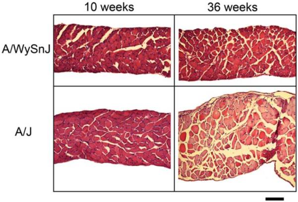
Muscle sections from diaphragms of young and mature A/J and wildtype (A/WySnJ) mice. Overt pathology is evident at 36 weeks of age, including rounded prenecrotic fibers, fatty infiltration, and central nuclei, whereas diaphragms from 10-week-old A/J mice appear relatively normal. Scale bar = 100 μm. [Color figure can be viewed in the online issue, which is available at www.interscience.wiley.com.]