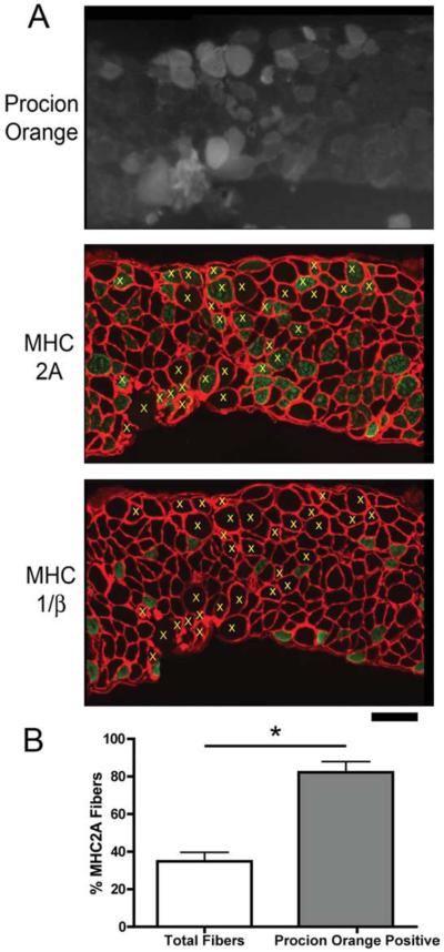
FIGURE 6.
MHC 2A fibers are most susceptible to membrane rupture in the diaphragms of A/J mice. (A) Procion orange uptake (upper panel), and MHC 2A and laminin immunohistochemistry (lower panel) in serial sections from a diaphragm of an A/J mouse. Yellow x's in the lower panel indicate Procion orange-positive fibers that are above background fluorescence, and show that most of the Procion orange fibers are positive for MHC 2A. Scale bar = 100 μm. (B) Quantification of MHC 2A fibers in n = 5 diaphragms show that less than half of all fibers are positive for MHC 2A, whereas more than 80% of the Procion orange fibers are positive for MHC 2A. [Color figure can be viewed in the online issue, which is available at www.interscience.wiley.com.]

