Figure 3.

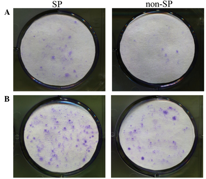
Colony formation of SP and non-SP cells in plates initially seeded with (A) 500 and (B) 1,000 cells (crystal violet staining). SP, side population.

Colony formation of SP and non-SP cells in plates initially seeded with (A) 500 and (B) 1,000 cells (crystal violet staining). SP, side population.