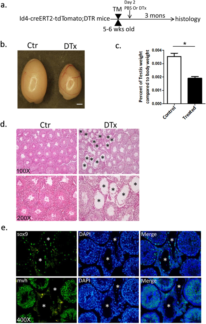
Figure 5. Ablation of Id4+ cells in mice results in a disruption of spermatogenesis.
(a) Experimental strategy for DTx treatment. (b) Intact testes from PBS-treated (Ctr) and DTx-treated mice (DTx). Id4-CreERT2-tdTomato;ROSA26-flox-stop-DTR mice were treated with PBS or DTx after TM induction. The testes of Id4-CreERT2-tdTomato;ROSA26-DTR mice appeared smaller in size after 3 months of DTx treatment. Scale bar, 1 mm. (c) Comparison of testes weights between control mice (Ctr) and DTx-treated Id4-CreERT2-tdTomato;ROSA26-DTR mice (DTx) (n = 7). *Denotes significant difference at P = 1.01133E-05. Statistical significance was determined using a two-tailed Student’s t-test. (d) H&E staining of paraffin-embedded sections from the testis of Id4-CreERT2-tdTomato;ROSA26-flox-stop-DTR mice that received a single tamoxifen with PBS treatment(Ctr, left column) or with single DTx treatment(DTx,right column). There are more degenerated tubules (*) in DTx-treated mice (right column) than in control mice (left column). (e) Imunofluorescence staining for Sox9 and Mvh confirmed the almost complete absence of germ cells in some tubules of DTx-treated Id4-CreERT2-tdTomato;ROSA26-DTR mice with very few germ cells remaining. The Mvh-stained cells are germ cells. The cells stained with Sox9 are Sertoli cells. Original magnifications are as indicated. *degenerated tubules.

