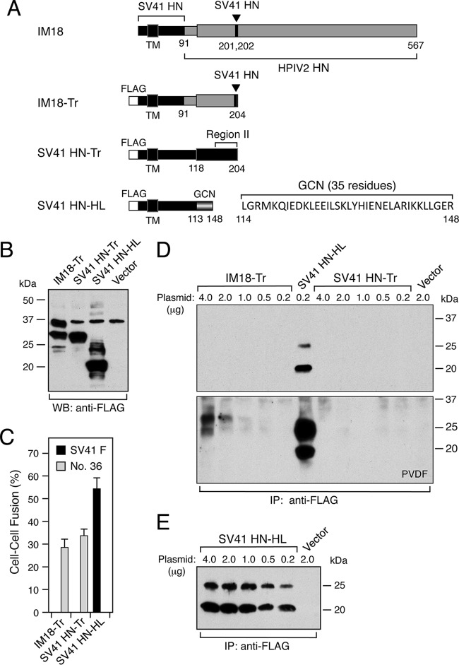
FIG 7.
Headless and truncated forms of the SV41 HN protein exhibit altered F protein specificity. (A) Schematic diagram of the HN proteins. FLAG, FLAG epitope tag; GCN, a modified terminal tetramerization module. (B) Detection of the HN proteins in the transfected cells. Subconfluent BHK cell monolayers in six-well culture plates were transfected with 2 μg/well of HN expression vector and lysed at 24 h posttransfection, and the cell lysate was subjected to SDS-PAGE under reducing conditions, followed by Western blotting with anti-FLAG monoclonal antibody. (C) F-triggering activity of the HN proteins. The average fusion index was determined as described in the legend for Fig. 1B; error bars indicate standard deviations. (D and E) Detection of cell surface-localized HN proteins. Cell surface biotinylation, immunoprecipitation with anti-FLAG monoclonal antibody, and SDS-PAGE analyses were performed as described in the legend to Fig. 3, except that 0.2 to 4.0 μg/well of expression vector was used for transfection and that PVDF membrane was employed for electroblotting in addition to the nitrocellulose membrane.

