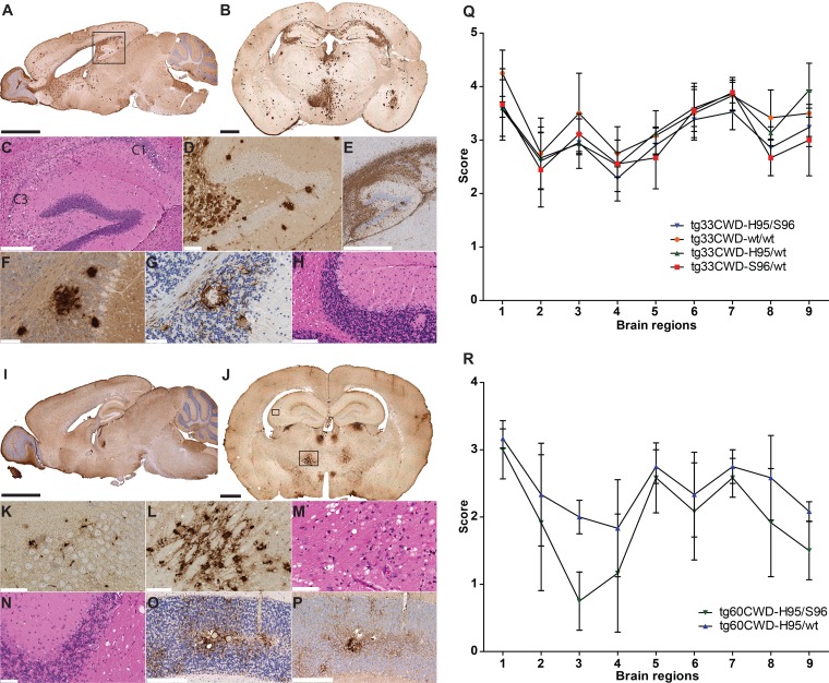
FIG 2.
Neuropathology of tg-deer-PRNP mice following the first passage of white-tailed deer CWD allotypes. (A and B) Accumulation of wt-PrPCWD aggregates in tg33 mice. The regional distribution of wt-PrPCWD aggregates was similar in mice receiving different CWD inocula (Fig. 3A to G). (C to E) Hippocampal degeneration (box in panel A) was characterized by spongiform change and a loss of pyramidal neurons of the Ammon's horn (C1 and C3), accompanied by the extensive accumulation of PrPCWD aggregates and abundant astrocytosis (GFAP). (F and G) Cerebellum pathology involved the loss of granular neurons and the presence of prion protein deposits flanked by astrocytes (as seen in sequential tissue sections). (H) Vacuolation was observed in Purkinje neurons and cerebellar white matter. (I and J) Detection of S96-PrPCWD aggregates in tg60 mice infected with the H95/wt and H95/S96 CWD allotypes. The distribution of PrPCWD aggregates was similar between animals receiving the H95+ deer CWD agent (Fig. 3J to K). (K) S96-PrPCWD aggregates in the hippocampus were noticeable at a higher magnification of the small box in panel J. (L and M) Abnormal prion protein deposits and spongiosis in thalamic nuclei shown by a higher magnification of the large box in panel J. (N to P) Cerebellar pathology included white matter vacuolation (N) and astrocytosis (O) that colocalized with diffuse and punctate protein aggregates (P). (Q) Lesion profile of tg33 mice infected with deer CWD allotypes. (R) Lesion profile of tg60 mice infected with the H95+ deer CWD agent. Brain regions are as follows: 1, medulla; 2, cerebellum; 3, superior colliculus; 4, hypothalamus; 5, thalamus; 6, hippocampus; 7, septum; 8, posterior cortex; 9, anterior cortex. Bars, 2.5 mm (A and I), 1 mm (B and J), 850 μm (E), 300 μm (C, O and P), 125 μm (D, L and H), and 60 μm (F, G, K, M, and N). PrPCWD detection was achieved with anti-PrP monoclonal antibody BAR224. (A) Brain section from a tg33 mouse infected with S96/wt CWD prions at 270 dpi; (B) brain section from a tg33 mouse infected with H95/S96 CWD prions at 387 dpi, (I) brain section from a tg60 mouse infected with H95/S96 CWD prions at 375 dpi; (J) brain section from a tg60 mouse infected with H95/S96 CWD prions at 414 dpi.

